This web page was produced as an assignment for Gen677 at UW-Madison Spring 2009
NFATc Interactions

Above is an example of a protein network, in humans, involving NFATc, which was obtained using STRING. A few familiar proteins can be seen in the diagram including CALN and DSCR1 which were both discussed as to their effects on NFATc in the Arron et al. paper discussed in the Scientific Article Review. The colored lines represent how the interaction between NFATc and the respective protein were discovered. Light-blue indicates a database search, yellow-green lines indicate textmining, and purple line relationships were found through experimental evidence. The only protein in this network that relates directly to NFATc that was found through experimental evidence is the Mxi2 protein, also known as mitogen-activated protein kinase 14. In the experiment it was found that NFATc is phosporylated by Mxi2 allowing it to accumulate in the nucleus. This is not dissimilar to the relationship between CALN and NFATc, which in not surprising as NFATc is a transcription factor and is most likely regulated and phosporylated by many proteins under different conditions. Other proteins contained in this network include other kinsases, phosphatases, and two different interleukin precursors. The interleukin precursors are of particular interest because they are shown to be important in stimulating immune response. (1) Given that FK506 is used after organ transplant to reduce the immune system reponse by inhibiting NFATc (Chemical Genetics) it is not surprising that interleukin precursors would be known to interact with NFATc.
Mouse Interaction Network
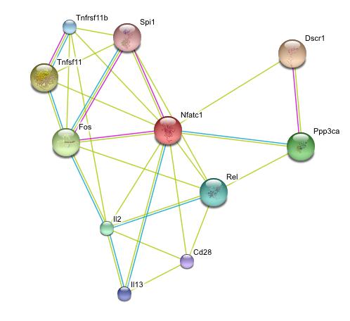
For comparison, the protein interaction of Nfatc for mice was also constructed using STRING. Here Dscr1 is found again along with Fos (an oncogene), a version of Ppp3c, and two interleukin proteins. Dscr1 is stilled described as a regulator of calcineurin, however a homolog for calcineurin is not shown in the diagram. However, Ppp3ca is a phosphatase that Dscr1 appears to regulate so it is possible that it may serve the calcineurin function in mice.
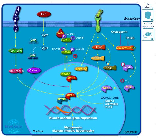
Control of Skeletal Myogenesis
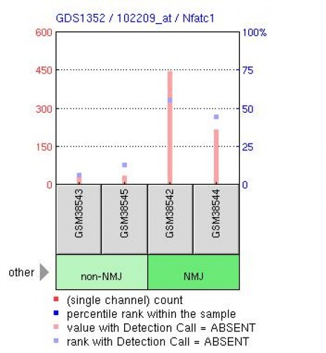
Expression Profile in Mouse
References:
1. Encyclopedia Britannica, Retrieved April 26, 2009 from http://www.britannica.com/EBchecked/topic/290335/interleukin
2. Biocarta. Control of skeletal myogenesis by HDAC & calcium/calmodulin-dependent kinase (CaMK). Retrieved May 13, 2009 from http://www.biocarta.com/pathfiles/h_hdacPathway.asp
Algorithmic Websites
STRING: http://string.embl.de/
BioCarta: http://www.biocarta.com/genes/index.asp
GEO: http://www.ncbi.nlm.nih.gov/geo/
*All images were obtained from the above websites
Margaret Noll, [email protected], last updated 5/13/2009/ http://www.gen677.weebly.com
